ADDED TO CART
Eye Anatomy: Parts Of The Eye
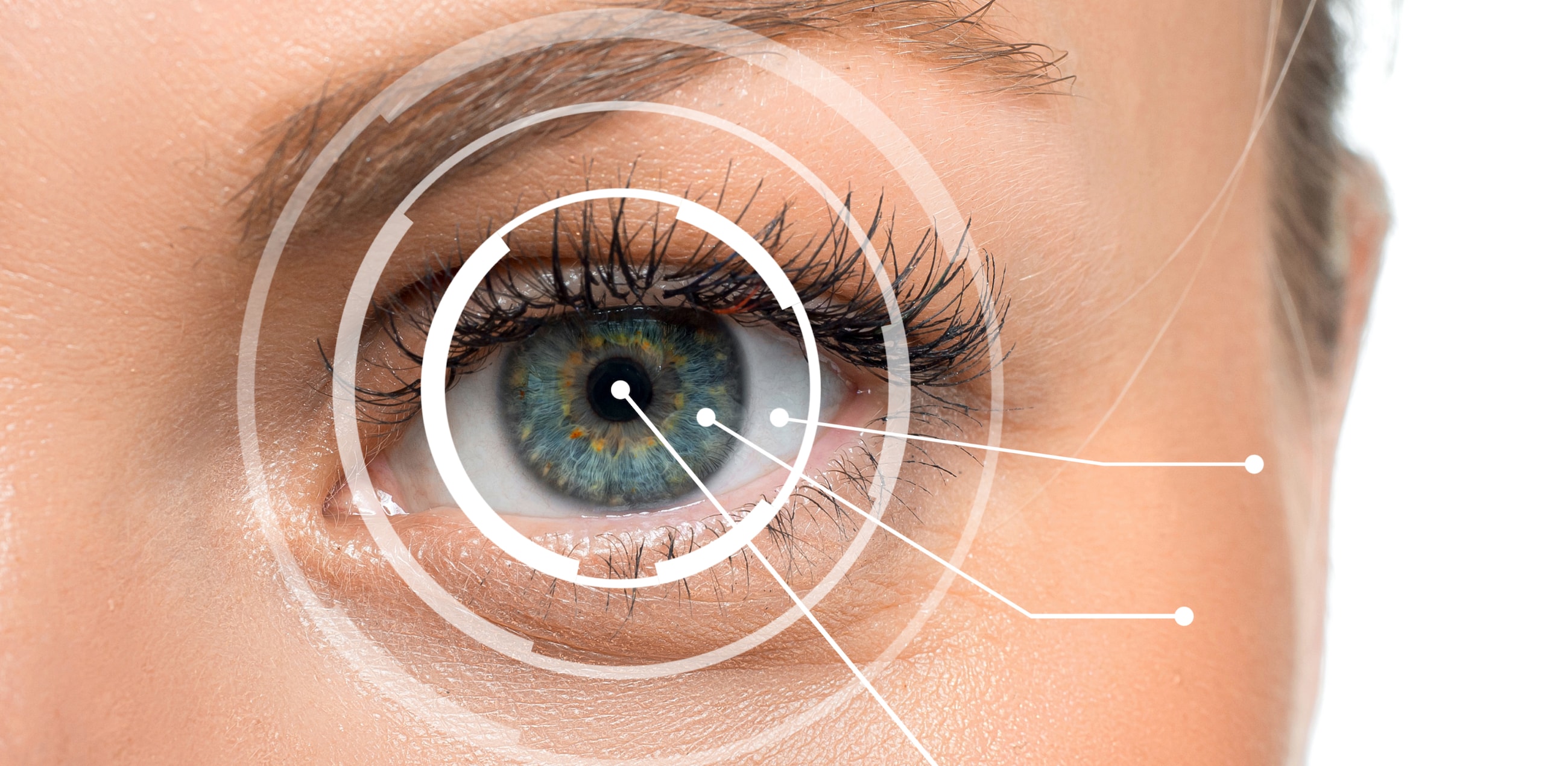
The eye's parts and layers, from front to back
Your eye is about 24mm across, roughly the size of a large
marble, yet that small space is packed with distinct working parts, and the
cornea, pupil, iris, retina and optic nerve each have their own job. Light
enters your eye, and the cornea and lens focus it so it lands right on your
retina, the eye's light-sensitive tissue. Signals from the retina travel to
your brain, which reads them as images.
Your eyes sit in the orbit, a bony socket cushioned by fat tissue. The orbit protects your eye and helps it work. It also holds the muscles and nerves that attach to the eyeball and keep your eyes moving in sync.
Six muscles move your eye in every direction. These are the extraocular muscles, and they attach all around the eyeball, pulling it up, down and side to side, and even rotating it.
Together, your two eyes give you depth perception and a field of view of around 200 degrees.
The eye makes the most sense in layers, working from the outer coat inward. The outer layer protects the eye and holds its shape. The middle layer feeds it and controls how much light gets in. The inner layer turns that light into the signals your brain reads as sight. In medical terms, the eyeball wall is built from three tunics, a tighter grouping than the outer, middle and inner layers below, since a few structures sit alongside the wall rather than inside it. The fibrous tunic is the wall's outer coat: the cornea and sclera. The vascular tunic, or uvea, is its middle coat: the iris, ciliary body and choroid. The nervous tunic is its inner coat, made of the retina. The lens, aqueous humor, conjunctiva and episclera don't belong to any tunic, though you'll still find them covered below.
Outer layers
The outer layers of the eye do several jobs, and one of the most important is protection.
Conjunctiva
The conjunctiva covers the inside of your eyelids, then wraps back over the white part of the eye. This is why it's impossible for contact lenses to get lost inside the eye! The conjunctiva folds back and becomes the outer covering of the white part (the sclera) of the eyeball.
It does more than keep contact lenses out of the eyeball. It also keeps the eye moist and lubricated and helps fight off infection. When someone gets "pink eye", conjunctivitis, the conjunctiva has become swollen and irritated, usually from an irritant, allergies or an infection.
Episclera
Just below your conjunctiva is the episclera. The prefix "epi" means above, and this layer lies above the sclera, which we cover next. It's packed with blood vessels that support your sclera, supplying oxygen and nutrition.
Sclera
When you see the whites of someone's eyes, you're looking at the sclera. It's a tough, fibrous layer that gives the eye its protection and its shape. The muscles that move the eyeball, those extraocular muscles, attach to it.
Cornea
The cornea at the front of your eye begins where the sclera ends. This clear tissue covers the colored part of your eye, the iris, and the opening where light enters, the pupil. It's only about 0.5mm thick at its center, thinner than a credit card, yet it does most of the work of focusing what you see.
The five-layered cornea handles most of the eye's focusing power thanks to its dome shape. Some people's corneas are shaped in a way that calls for astigmatism correction, either with glasses or toric contact lenses.
The cornea does more than focus light. It also acts as a barrier against particles and germs, and it's remarkably sensitive, its free nerve endings are 300 to 600 times denser than those in your skin! It even filters out a good deal of the sun's ultraviolet radiation.
Middle layers
The middle layers of your eye are busy, dynamic structures. They control how much light gets in, and they feed the inside of the eye oxygen and nutrients through a network of blood vessels.
Iris
What color are your eyes? That color comes from the iris. And if you've ever wondered why some eye colors are rarer than others, it comes down to how much pigment sits in this layer. The iris controls how much light reaches the back of the eye, and it's made of muscles that react to light by making your pupil larger or smaller.
Pupil
The dark circle inside the iris, the pupil, is where light enters your eye. In bright light, the iris shrinks the pupil down to about 2mm across; in dim light, it opens up to around 8mm, a fourfold change in size.
Lens
Your lens sits just behind the iris, so light coming through the pupil passes straight through it. This clear, crystalline lens fine-tunes the focus. It thickens when you look at something close up so the view stays sharp, a shift called accommodation.
Sometime in your mid-40s, the lens starts to stiffen, which brings on presbyopia. That's when most people begin to reach for bifocal and multifocal contact lenses and glasses. The lens can also grow cloudy with age, a common change many people first notice after 40.
Anterior chamber and posterior chamber
The anterior and posterior chambers sit in the front part of the eye, spanning from the cornea to the front of the crystalline lens. The space from the cornea to the iris is the anterior chamber. The space from the iris to the front of the lens is the posterior chamber.
Aqueous humor
A clear, watery fluid called aqueous humor is made continuously in the posterior chamber. It comes from a structure called the ciliary body, which sits behind the iris. The aqueous humor flows through the pupil into the anterior chamber, then drains out where the iris meets the cornea.
When the making and draining of aqueous humor fall out of balance, pressure inside the eye can build up. Doctors check for sustained high eye pressure during a comprehensive eye exam. Left unchecked over time, that pressure can affect the optic nerve, the nerve that carries visual signals from the eye to the brain.
Ciliary body and ciliary muscle
The ciliary body makes aqueous humor, and it also houses the ciliary muscle. That muscle is what thickens the crystalline lens when you look up close.
Choroid
This thin layer of blood vessels keeps the outer retina — the inner light-sensitive tissue at the back of the eye, nourished, delivering oxygen and nutrients and carrying away waste. It sits between the sclera and the retina, and it's part of the uvea. The uvea is made of the iris, the ciliary body and the choroid, the same middle, vascular layer named earlier.
Inner layers
Most of us take clear vision for granted, but your ability to see depends entirely on this innermost layer.
Vitreous humor
Between the back of the lens and the retina sits a substance that fills the back two-thirds of the eye: the vitreous humor. It's clear and jelly-like, and it supports the inside of the eye while holding its round shape.
Retina
The innermost lining at the back of your eye is the retina, a delicate sheet of tissue less than 0.5mm thick. It's home to cells called photoreceptors: rods, which pick up dim light and motion, and cones, which detect color and fine detail. Light travels through the cornea and lens and past the vitreous humor, then lands on the retina. There, it triggers the rods and cones, which turn the light into electrical signals.
Macula and fovea
The macula lutea is a patch of the retina with a very high concentration of photoreceptor cells. It sits centrally, delivering detailed vision plus much of our color vision.
The fovea centralis is a tiny pit in the macula, packed with cones. It gives us our sharpest vision of all.
Optic nerve
Once the photoreceptors turn light into electrical signals, nerve fibers carry those signals to the brain. Those fibers bundle together to form the optic nerve. More than one million nerve fibers make up a single optic nerve.
The brain reads the electrical signals coming down the optic nerve as images, and you experience all the shapes and colors of the world around you!
Every layer covered here leans on the ones beside it. That's why a comprehensive eye exam checks far more than how clearly you can read a chart. It's worth booking one every year, even when your vision still feels sharp. Once your prescription is confirmed, most vision plans and FSA/HSA benefits help cover the contacts that match it.